FIG 4.
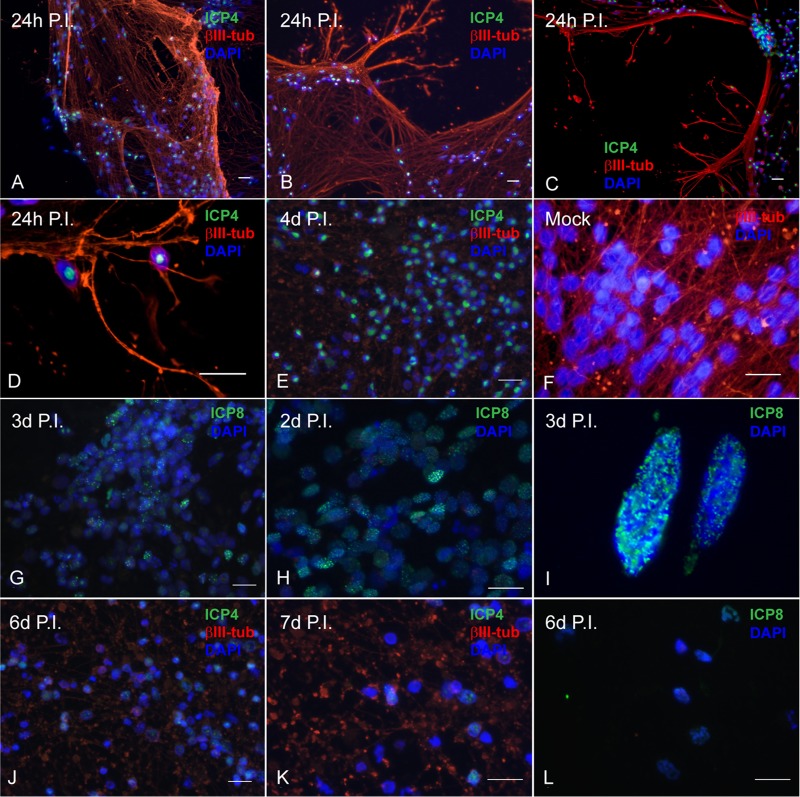
Lytic HSV-1 proteins are abundantly expressed in LUHMES neuronal cultures following acute infection and are reduced during latency. Differentiated, postmitotic LUHMES neurons were infected with 17syn+ at an MOI of 3 and analyzed by immunofluorescence for expression of viral lytic proteins over a time course of 7 days. (A to E) During the acute infection, ICP4 (shown in green) is abundantly expressed in almost all the neurons as large well-developed centers in the nucleus (1 to 4 days p.i.). (F) Mock-infected LUHMES cells. (G to I) Punctate, nuclear, chromatin-associated ICP8 (shown in green) is also evident during the acute infection (2 to 3 days p.i.). (J to L) By 6 to 7 days p.i., a dramatic reduction in both ICP4 (J and K) and ICP8 (L) expression is seen. Scale bars represent 20 μm. Neurofilaments are stained for βIII-tubulin (red); nuclei are stained with DAPI (blue).

