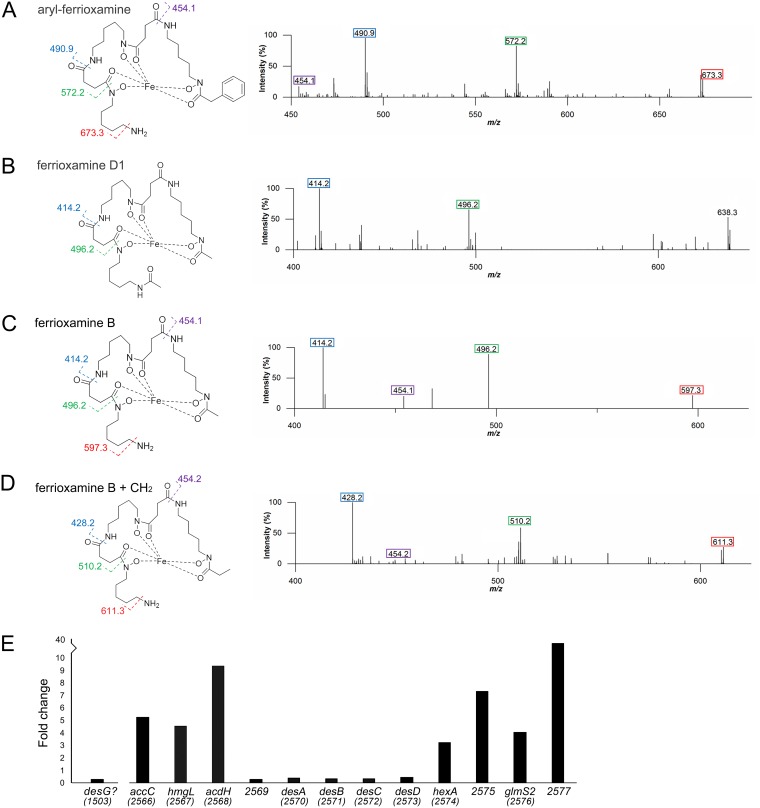
FIG 3.
Explorer cells produce ferrioxamines. Panels A to D show accurate mass fragmentation (MS2) data and molecular structures of associated ferrioxamine (iron-complexed desferrioxamine) compounds. Fragments that are common to this class of molecules have been indicated with colored boxes on each molecule, along with colored numbers above the associated fragment in the spectra. (A) Aryl-ferrioxamine, from S. venezuelae exploring on YP and S. venezuelae exploring beside S. cerevisiae on YPD. (B) Ferrioxamine D1, from S. venezuelae exploring on YP and S. venezuelae exploring beside S. cerevisiae on YPD. (C) Ferrioxamine B, from S. venezuelae exploring on YP and S. venezuelae exploring beside S. cerevisiae on YPD. (D) Ferrioxamine B plus CH2 (where the CH2 is depicted as an ethyl group, as the methylene addition is on the right-hand side of the molecule and is most likely located in the acyl tail) from S. venezuelae exploring beside S. cerevisiae on YPD. (E) Normalized transcript levels for desG and genes in the S. venezuelae desferrioxamine biosynthetic cluster (as defined by antiSMASH [20]) in explorer cells, divided by those determined for nonexploring cells. The associated gene names or sven gene numbers are shown below each gene.

