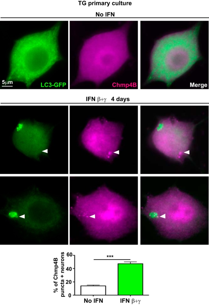
FIG 7.

IFNs induce Chmp4B puncta in TG neurons. Representative images from immunofluorescence microscopy of TG neurons from LC3-GFP+/− mice in culture not treated or treated with IFN β+γ for 4 days. LC3-GFP is shown in green and Chmp4B in magenta. White arrowheads point to Chmp4B puncta. Graph shows quantification of neurons containing Chmp4B-positive puncta in the cultures; n = 8 (>400 neurons were counted for each condition). The experiment shown is representative of two independent experiments. ***, P < 0.001.
