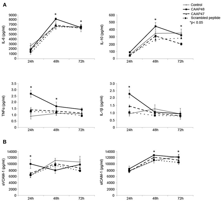
Figure 6.
Release of cytokines and adhesion molecules in the liver-on-chip model stimulated with CAAP48. (A) Cytokine concentrations of IL-1β, IL-6, TNFα, and IL-10 within the supernatants of the vascular layer were determined by flow cytometry using a bead-based multiplex immunoassay. (B) The release of sVCAM-1 and sICAM-1 within the supernatants of the vascular layer were examined by Bio-Plex assay. Results of six independent experiments are shown.

