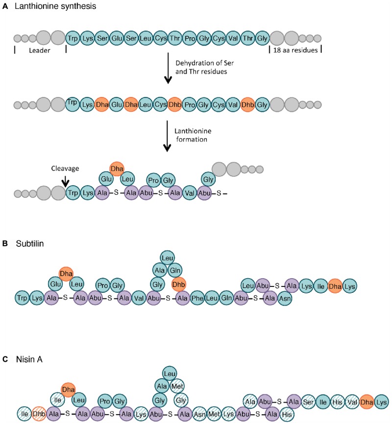
Figure 3.

Lanthionine biosynthesis. General pathway of the lanthionine synthesis (A), structure of subtilin (B) and nisin A (C). Non-modified AA are indicated in teal whereas dehydrated serine (Dha, dehydroalanine) and threonine (Dhb, dehydrobutyrine) are colored in orange. The lanthionine (Ala-S-Ala, alanine-S-alanine) and R-methyllanthionine (Abu-S-Ala, aminobutyrate-S-alanine) bridges are shown in purple. The AA of nisin that differ from those in subtilin are highlighted as hatched circles. Adapted from Cotter et al. (2005) and Spieß et al. (2015).
