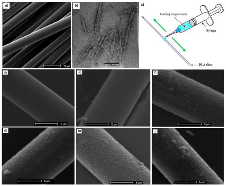
Figure 5.
(a) Scanning electron microscopy (SEM) image of PLA fibers obtained at 400 m min−1; (b) Transmission electron microscopy (TEM) of cellulose nanocrystals; (c) schematic of the coating procedure employed on the PLA fiber surface; and SEM images of: (d) noncoated; (e) PLA/PVAc; (f) PLA-CNCs-65; (g) PLA-CNCs-75; (h) PLA-CNCs-85; and (i) PLA-CNC-95 fibers. Reprinted from [84] Copyright ©2018 American Chemical Society.

