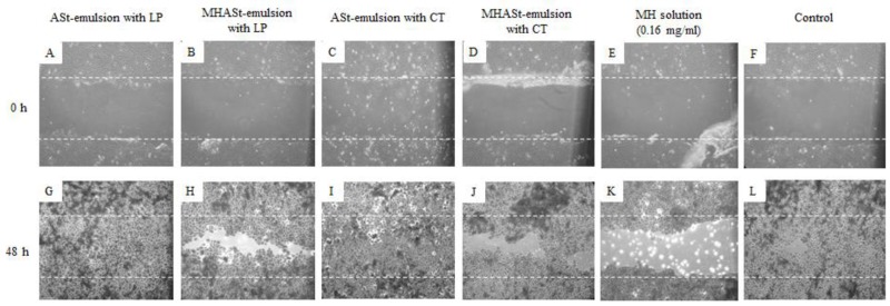
Figure 4.
Representative time-lapse images of HaCaT keratinocyte scratch assays immediately after the scratches had been made and then after 48 h in the presence of 5%-emulsions with LP (A,G), 5%-MHASt-emulsions with LP (B,H), 5%-ASt-emulsions with CT (C,I), 5%-MHASt-emulsions with CT (D,J), MH solution (E,K) or control medium (F,L). The cells were allowed to migrate for 48 h, fixed and photographed. Outlines of the original wounds are marked with dashed lines. Original magnification 40×.

