
FIGURE 5.
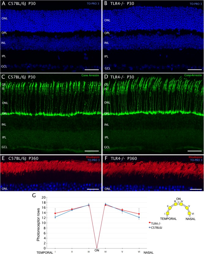
Number and morphology of photoreceptors in control and TLR4-deficient mice. (A–F) Vertical sections from control (A,C,E; C57BL/6J) and TLR4-deficient (B,D,F; TLR4–/–) mice retinas stained with the nuclear marker TO-PRO 3 (blue) to visualize all cell nuclei (A,B,E,F), and labeled for cone arrestin (green) to visualize cone photoreceptors (C,D) or for rhodopsin (red) to evidence rod outer segments (E,F). (G) Quantitation of photoreceptor rows in the ONL in both C57BL/6J and TLR4–/– retinas (n = 5 in both cases). The scheme to the right of the panel shows the position of each representative region analyzed in the retina. Error bars represent the SEM. OS, outer segment; IS, inner segment; ONL, outer nuclear layer; OPL, outer plexiform layer; INL, inner nuclear layer; IPL, inner plexiform layer; GCL, ganglion cell layer; ON, optic nerve. Scale bar: 40 μm.