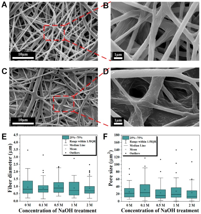
Figure 3.
Surface morphology of PCL and hydrolyzed electrospun nanofibers. Compared to electrospun PCL nanofibers (A,B), hydrolyzed electrospun PCL nanofibers (C,D) after 1.5 h of 2 M NaOH treatment shows slight degradation at the external fibers while flattening and denser arrangement in the internal. The fiber diameter (E,F) pore size distributions of the electrospun PCL nanofibers at different concentrations of NaOH treatment shows no significant differences at the mean values (p > 0.05), n = 47 for each concentration in (E,F), respectively.

