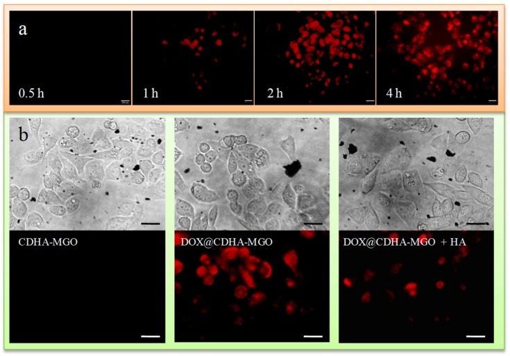
Figure 6.
(a) Photomicrographs of BEL-7402 cells treated with DOX@CDHA–MGO (20 μg·mL−1) at 37 °C for incubation times of 0.5, 1, 2, and 4 h, observed under an inverted fluorescence microscope. (b) Fluorescence microscopy images of BEL-7402 cells incubated for 2 h at 37 °C with CDHA–MGO and DOX@CDHA–MGO in the presence or absence of free HA. Scale bar = 20 μm.

