Figure 1.
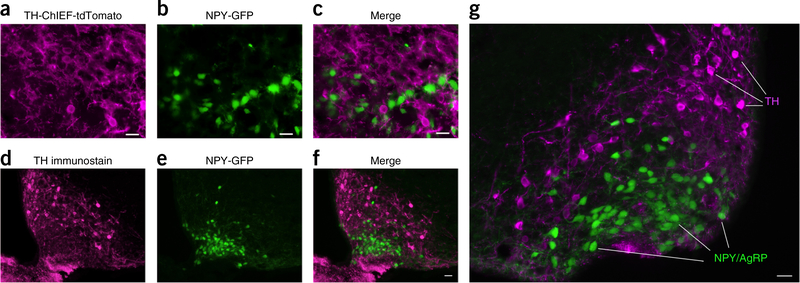
Lack of colocalization of TH in NPY/AgRP neurons. (a) TH-ChIEF-tdTomato neurons are shown in magenta after Cre-dependent AAV-ChIEF-tdTomato (arrows) was injected into ARC of a TH-Cre mouse crossed with an NPY-GFP mouse. (b) NPY-GFP neurons are shown in the ARC on the same section as in a. (c) Merged image shows that TH neurons do not overlap with NPY/AgRP cells. Scale bar, 20 μm. We analyzed 45 sections from 3 mice with a combination of low and high magnification fluorescence microscopy; 1,561 TH-ChIEF-tdTomato expressing cells were identified and none expressed the NPY/AgRP GFP. Similarly, 2,756 NPY/AgRP-GFP expressing cells were examined and none expressed detectable TH-ChIEF-tdTomato. (d) Arcuate TH immunostained cells are shown in magenta from an NPY-GFP transgenic mouse. (e) Green neurons are shown from the same section of the NPY-GFP transgenic mouse in d. (f) Merged image reveals no overlap between magenta TH-ChIEF-tdTomato and green NPY/AgRP neurons. Scale bar, 20 μm. (g) Higher magnification of a similar merged image is shown from another section. 18 sections from 3NPY-GFP mice were examined. Scale bar, 20 μm.

