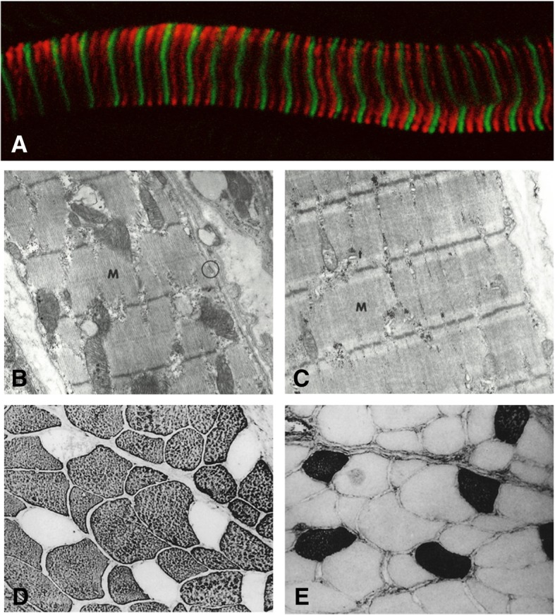
Fig. 4.

a Longitudinal section of rat extraocular muscle stained with MYH7b (red) and α-actinin (green) adapted with permission of the publisher from Rossi et al. 2010, copyright©2010 John Wiley and Sons. b, c Electron micrograph of muscle spindle chain fiber (b) characterized by clearly defined sarcomeres and large mitochondria and bag fiber (c) characterized by less well-defined sarcomeres and fewer mitochondria Reprinted with permission of the publisher from Ovalle et al. 1971, ©1969, CCC Republication. d, e Immunoperoxidase staining of MyHC-masticatory (d) and β-MyHC (e) in cat masseter muscle sections shows the majority of fibers are comprised of MyHC-masticatory and only a small proportion of fibers express β-MyHC, in contrast to humans, which express β-MyHC as the predominant isoform. Reprinted with permission of the publisher from Kang et al. 2010, ©2010, SAGE Publications
