Fig. 1.
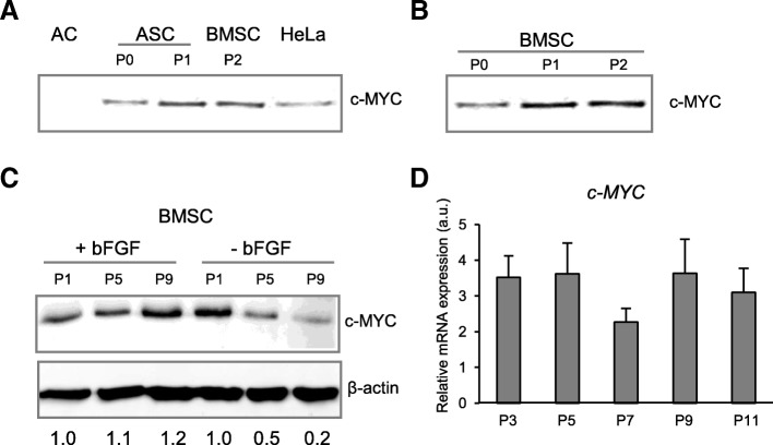
c-MYC protein accumulation that was increased upon ex vivo passaging. a, b Western blot analysis of c-MYC protein abundancies in different human mesenchymal cell types. Cellular extracts from 25,000 cells for every sample were analyzed by Western blot, with equal volume loading. a c-MYC was assayed in freshly isolated articular chondrocytes (AC), MSC from adipose tissue (ASC), bone marrow MSC (BMSC), and Hela cells served as a positive control for c-MYC protein accumulation. b Bone marrow-derived MSC isolated at passages 0 (P0), 1 (P1), and 2 (P2). c, d c-MYC protein accumulation and mRNA expression were monitored by Western blot (C) and qRT-PCR (n = 3) (d) in BMSC at indicated passages (P1, P3, P5, P7, P9, P11), in media with or without basic fibroblast growth factor (bFGF), as indicated (c); numbers below WB in C indicate semi-quantitative evaluation of c-MYC protein abundancies normalized to β-actin, as a fold change to a corresponding P1

