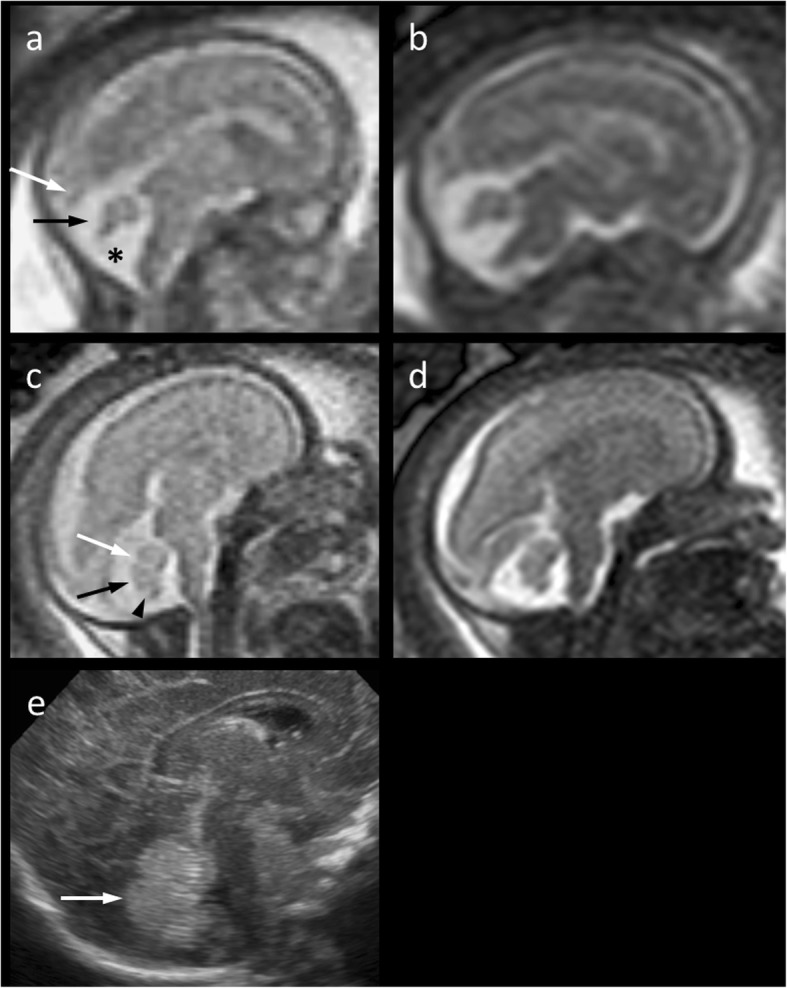
Fig. 1.

a–d Mid-sagittal T2-weighted 1.5 Tesla magnetic resonance images (a and c, Single-shot; b and d, Balanced Turbo Field Echo) of the fetal brain. At 21 weeks‘gestation (a, b), the cerebellar vermis (a, black arrow) appears upwardly rotated and moderately hypoplastic with a normal torcular position (a, white arrow). At 27 weeks‘gestation (c, d), the vermis appears nearly normal in position, shape and size suggesting delayed fenestration of Blake’s pouch (a, asterisk) The primary (c, white arrow), prepyramidal (c, black arrow), and secondary (c, arrow head) fissures are roughly discernible. e A slight uncertainty regarding minimal hypoplasia of the cerebellar vermis (arrow) remains even after inconspicuous transcranial ultrasound postnatally at the age of 10 weeks
