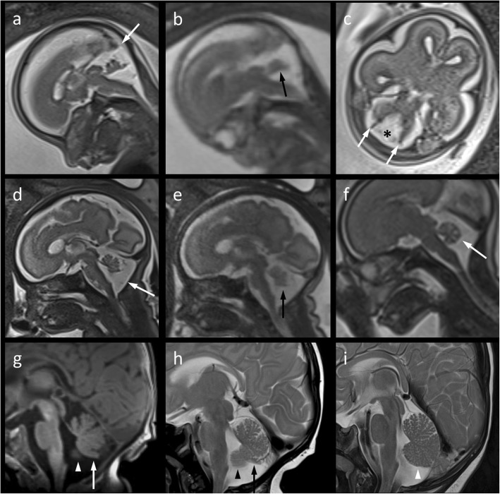
Fig. 2.
a–c Mid-sagittal (a, Half-Fourier Acquisition Single-Shot Turbo Spin-Echo [HASTE]; b, True Fast Imaging With Steady-State Free Precession) and axial (c, HASTE) T2-weighted 3 Tesla magnetic resonance (MR) images of the fetal brain at 21 weeks‘gestation suggesting a moderately hypoplastic cerebellar vermis with a flattened fastigial point (b, arrow) and moderately increased tegmento-vermian angle of about 35°, but normal torcular position (a, arrow). Lateral septa (c, arrows) in the posterior fossa are believed to belong to Blake’s pouch (c, asterisk). d, e Follow-up imaging on the same scanner at 31 weeks‘gestation shows nearly normal rotation of the vermis in a slightly enlarged posterior fossa (d, arrow) suggesting delayed fenestration of Blake’s pouch. Mild infero-posterior vermian hypoplasia (e, arrow) may be suspected from early third-gestational MR imaging (MRI) (d, e) This finding becomes more evident if compared to the sagittal T2 HASTE image of the cerebellar vermis (f, arrow) in a fetus at 24 weeks‘gestation scanned for suspected pulomonary sequestration on the same MR system. g, h A similar pattern is depicted by postnatal sagittal MRI (g, T1 Magnetization-Prepared Rapid Gradient-Echo; h, T2 Turbo spin echo) at the age of 12 weeks. Size and shape of the cerebellar vermis imply that it is mildly hypoplastic and its posterior lobe has experienced mass effect due to prolonged persistence of Blake’s pouch. Further, partial volume of cerebellar hemisphere (g, h; arrow heads) adjacent to the foramen of Magendie has to be considered. i For comparison of cerebellar volume and fissuration, the sagittal T1-weighted MR image in a 4-month-old, clinically inapparent infant scanned for a temporopolar arachnoid cyst – known from pre- and postnatal ultrasound – is given here

