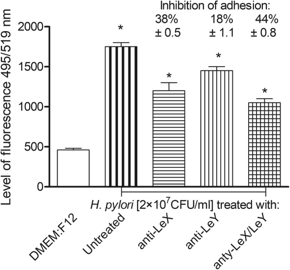
Fig. 3.

Involvement of H. pylori Lewis (Le) X or LeY determinents in adhesion to guinea pig primary gastric epithelial cells. Binding of H. pylori to gastric epithelial cells was evaluated by imaging H. pylori stained with anti-H. pylori antibodies (Ab) conjugated with fluoresceine isothiocyanate (FITC). Gastric epithelial cells were cocultured for 2 h with live H. pylori (2 × 10⁷ colony forming units –CFU/ml) non treated or treated for 30 min with anti-LeX, anti-LeY or both types of antibodies. The intensity of fluorescence was measured in a fluorescence reader at 495 nm excitation/519 nm emission, mean values ±SD. * Statistical significance for cells exposed to H. pylori untreated with anti-LeX or anti-LeX and anti-LeY antibodies, vs cells exposed to H. pylori, treated such antibodies, p < 0.05 in the non parametric U Mann-Whitney test
