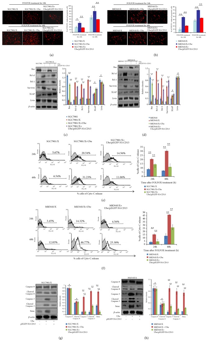
Figure 3.
Ubenimex regulates the expression of apoptosis-related proteins to promote FOLFOX-induced apoptosis in GC cells by suppressing CD13 expression. ((a) and (b)) Indicted cells were pretransfected with pEGFP-N1-CD13 and then treated with Ubenimex; evaluation of FOLFOX-induced apoptotic SGC7901/X (a) and MKN45/X (b) cells was confirmed by TUNEL staining of red fluorescent (upper panels); the proportion of apoptotic cells was also shown as means ±SD from three independent experiments (bottom panels). ∗P < 0.05 and ∗∗P <0.01. ((c) and (d)) Expressions of CD13 and apoptosis-related proteins in SGC7901/X (c) and MKN45/X (d) cells were determined via western blotting analysis. Data are also shown as representatives (left panels) and mean ±SD relative gray values normalized to β-actin expression from three independent experiments (right panels). ∗∗P < 0.01 and #P>0.05. ((e) and (f)) Release concentration of Cyto-C in the culture supernatant of FOLFOX-treated SGC7901/X (e) and MKN45/X (f) cells with the treatment of Ubenimex or/and pEGFP-N1-CD13 plasmid, was measured by flow cytometric analysis (left panels). Data are also expressed as means ± SD of positive cell numbers from three independent experiments (right panels). ∗∗P<0.01. ((g) and (h)) Indicated cells were treated with the combination of Ubenimex and pEGFP-N1-CD13 plasmid for 24h and then stimulated with FOLFOX regimen for another 24h. Protein levels of Smac, total caspase-3 and caspase-8, or cleaved caspase-3 and cleaved caspase-8 in SGC7901/X (g) and MKN45/X (h) cells were detected by western blotting analysis. Data are shown as representatives (left panels) or means ± SD from three independent experiments (right panels). ∗∗P < 0.01 and #P>0.05.

