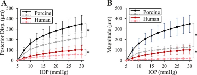
Figure 4.

(A) Posterior displacement of the ONH (solid lines) and PPT (dashed lines) in human donor eyes (n = 14) and porcine eyes (n = 12), and (B) posterior displacement of the ONH (solid lines) and scleral canal expansion (dashed lines) in human donor eyes (n = 14) and porcine eyes (n = 12). Asterisk (*) indicates statistical significance (P < 0.05) in paired t-tests at 30 mm Hg.
