Figure 4.

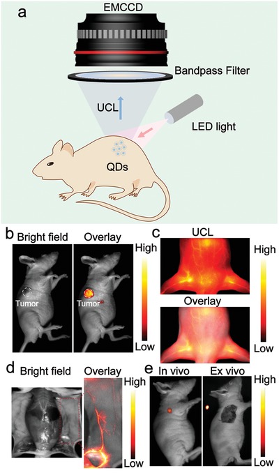
a) Schematic diagram of low‐power LED light excited bioimaging in vivo. A 980 nm LED light (20 mW cm−2) excited b) tumor, c) blood vessel in vivo, d) blood vessel in situ, and e) lymph node UCL imaging of UCL‐QDs.

a) Schematic diagram of low‐power LED light excited bioimaging in vivo. A 980 nm LED light (20 mW cm−2) excited b) tumor, c) blood vessel in vivo, d) blood vessel in situ, and e) lymph node UCL imaging of UCL‐QDs.