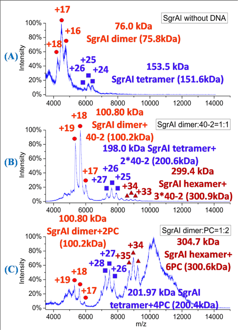
Figure 1.

Spectra of SgrAI and SgrAI with different DNA. (A) SgrAI without DNA. Peaks corresponding to the SgrAI dimer (red circles) and SgrAI tetramer (purple squares) are visible. (B) SgrAI with 40–2 DNA (molar ratio 1:1). Peaks corresponding to the SgrAI dimer bound to one duplex of 40–2 DNA (red circles), SgrAI tetramer bound to two 40–2 duplexes (purple squares), and SgrAI hexamer bound to three 40–2 duplexes (red triangles) are visible. (C) SgrAI with PC DNA (molar ratio 1:2). Peaks corresponding to the SgrAI dimer bound to two PC DNA duplexes (red circles), SgrAI tetramer bound to four PC DNA duplexes (purple squares), and SgrAI hexamer bound to 6 PC DNA duplexes (red triangles) are visible, along with a large unresolved peak (900–1200 m/z). The theoretical masses were listed in the parentheses.
