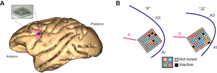
Figure 2.
Neurophysiological recordings. A, Location of chronically implanted multielectrode Utah array within the left caudal LPFC. The shaded pink area roughly represents area 8A in the macaque brain. The blue square represents implant location. P: principal sulcus. AS: arcuate sulcus superior. AI: arcuate sulcus inferior. B, Implant location based on intra-operative photography for both monkey “F” and monkey “JL” in reference to major sulci. Each small square represents one of the 96 microelectrodes on the array. Colors represent the spatial attentional tuning of the neurons recorded at each electrode site as a function of the four quadrant locations (inset). Note tuned stands for neurons that do not show attentional modulation. Inactive represents reference electrodes and grounds.

