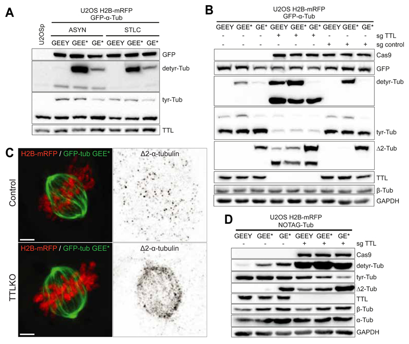
Fig. 4.
A. Western-blot analysis of U2OS cell lysates stably expressing H2B-mRFP and GFP-α-Tub (GEEY, GEE* or GE*) in asynchronous and mitotic populations (cells treated with STLC for 14h). B. Western-blot analysis of U2OS cell lysates stably expressing H2B-mRFP and GFP-α-Tub (GEEY, GEE* or GE*) in control (sgcontrol) and TTL KO cells (sgTTL). C. Deconvolved immunofluorescence showing the cellular distribution of ?2-tubulin in U2OS cells stably expressing H2B-mRFP and GFP-GEE* in the presence or absence of TTL. Scale bar, 5 μm. D. Western-blot analysis of U2OS cells lysates stably expressing H2B-mRFP and NOTAG-α-Tub (GEEY, GEE* or GE*), in control and TTLKO (sgTTL) cells.

