Fig. 1.
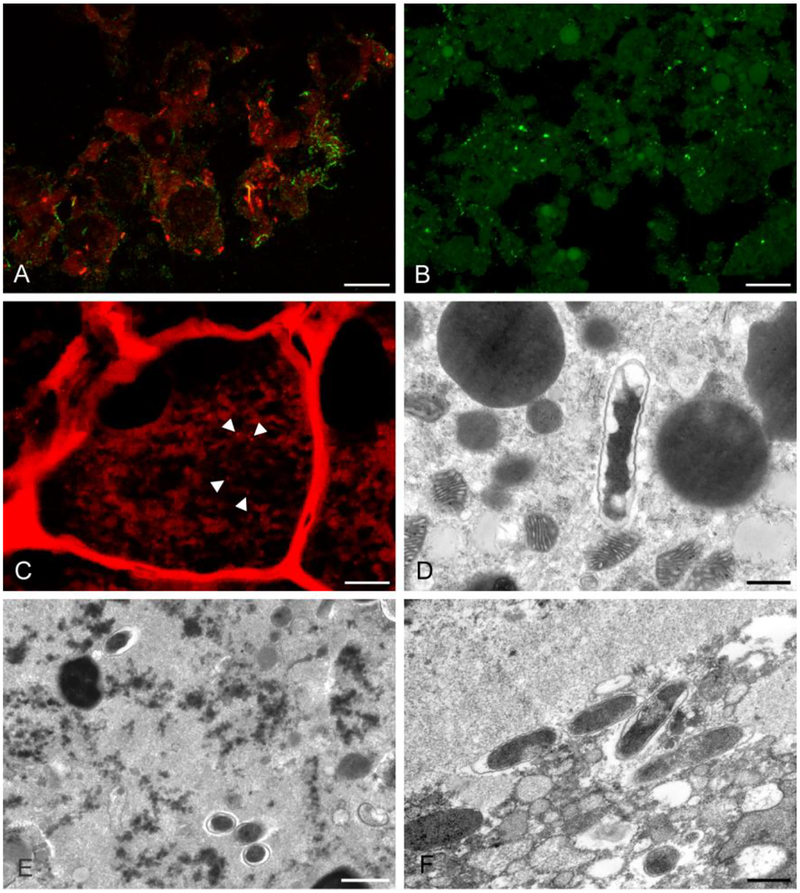
Immunohistochemistry (IHC) and fluorescence in situ hybridization (FISH) [A-C] and transmission electron microscopy (TEM) [D-F] revealed rickettsial organisms in the experimentally infected tick tissue. (A) R. parkeri (green) and “Candidatus R. andeanae” (orange) were detected in Vero cells (FISH and IHC, scale bar = 10 μm). (B) R. parkeri (green) were detected in salivary gland in the co-infected group on DPT 12 (IHC, scale bar = 10 μm). (C) “Candidatus R. andeanae” (orange) were detected in developing eggs in an ovary from the co-infected group on DPT 12 (FISH, scale bar = 10 μm). (D) A single rickettsial organism in a developing egg in the ovary of a “Candidatus R. andeanae” singly-infected tick from DPT 12 (TEM, scale bar = 600 nm). (E) Rickettsial organisms in midgut tissue from a tick in the co-infected group sampled on DPT 12 (TEM, scale bar = 1 μm) (F) Multiple rickettsial organisms in salivary gland tissue from a tick in the co-infected group sampled on DPT 12 (TEM, scale bar = 600 nm).

