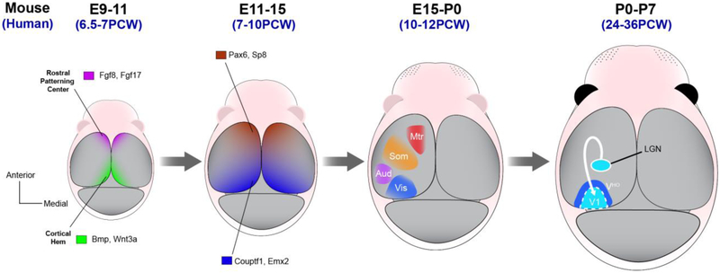
Figure 1. Patterning the cortical area map.
Illustrations of the dorsal surface of the developing mouse brain at different pre and postnatal stages. The first stage depicts the morphogen signals arising from the rostral patterning center (purple, Fgf8 and Fgf17) and cortical hem (green, Bmp and Wnt) beginning around embryonic day 9 in mice (corresponds approximately to human gestational week 6 or 7). These morphogens induce gradients of transcription factor (TF) expression, including Sp8, Pax6, Emx2 and Couptf1. The transcriptional programs regulated by these transcription factors establish cortical fields and the guidance cues that attract area-specific thalamic innervation. The innervation of cortex by thalamic axons, which happens postnatally in rodents (this occurs in the second and third trimester of human pregnancy), drives the sharpening of molecular and cytoarchitectural boundaries between primary sensory cortex (e.g. V1, dark blue) and adjacent higher order cortical areas (e.g. VHO, light blue).

