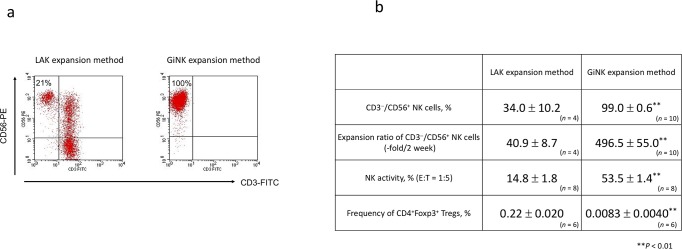
Fig 1. Characteristics of expanded cells from human PBMCs in different culture methods.
(a) Representative flow cytometric figures depicting the frequency of CD3−CD56+ NK cells in expanded cells. The cells were stained with FITC-conjugated anti-CD3 and PE-conjugated anti-CD56. (b) The profile of cells expanded by two different culture methods. The figure shows the purity of NK cells (%), the expansion rate of NK cells (fold), the NK activity (%), and the frequency of Tregs (CD4+Foxp3+ cells, %) in two culture methods. Data indicated as mean ± SE. P values were determined using the Mann–Whitney U test to compare NK cells expanded by the newly established method and LAK expansion method. Statistically significant differences: **P < 0.01.

