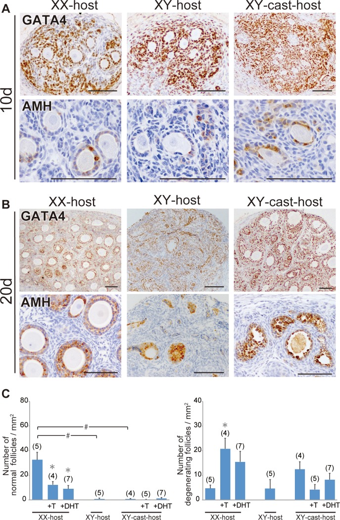
Fig 1. Repression of initial follicular growth in ovarian grafts under androgen-excess host conditions.
(A, B) Anti-GATA4 and AMH immunostaining of wild-type ovarian tissues grafted into intact male (XY-host), intact female (XX-host) and castrated male (XY-cast-host) hosts treated with or without testosterone (T), or dihydrotestosterone (DHT). The lower magnified images of GATA4-positive gonadal areas are shown in upper plates in A and B. AMH-positive healthy primary, secondary, and antral follicles were detected in ovaries grafted into female hosts (lower plate in B). (C) Bar graphs indicate the relative numbers of normal healthy follicles (left) and degenerating follicles (right). The data are expressed as means ± SEM (*p<0.05 as compared with non-treated host value in each host group; #p<0.05 as compared between two groups, Steel's test). Each number in parentheses indicates the number of explants used in each host. Scale bars, 100 μm for (A, B).

