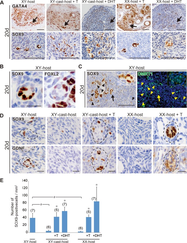
Fig 2. Ectopic appearance of SOX9-posititve Sertoli-like cells in ovarian grafts under androgen-excess host conditions.
(A–D) Anti-GATA4, SOX9, FOXL2, DMRT1, or GDNF immunostaining of wild-type ovarian tissues grafted into XY-, XX-, and XY-cast-hosts treated with or without T or DHT on days 20 post-transplantation. The lower magnified images of GATA4-positive gonadal areas are shown in upper plates in a. Normal follicular structures were mostly absent, and tubular structures containing ectopic SOX9-positive cells were evident near the edge of ovaries grafted into XY-host and T/DHT-treated XX- and XY-cast-hosts on day 20 post-transplantation (arrows in A). The ectopic SOX9-positive cells are FOXL2-negative (B), and they are found in the DMRT1-positive/GDNF-positive tubular region (C, D). In c, the inset shows high-magnification images of tubular structures indicated by arrows. (E) Bar graphs indicate the SOX9-positive cells per gonadal area (mm2). The data are expressed as means ± SEM (*p<0.05 as compared with non-treated host value in each host group; #p<0.05 as compared between two groups, Steel's test). Each number in parentheses indicates the number of explants used in each host. Broken lines indicate the border of tubular structures in c and e. Scale bars, 100 μm for (A, C); 20 μm for (B, D).

