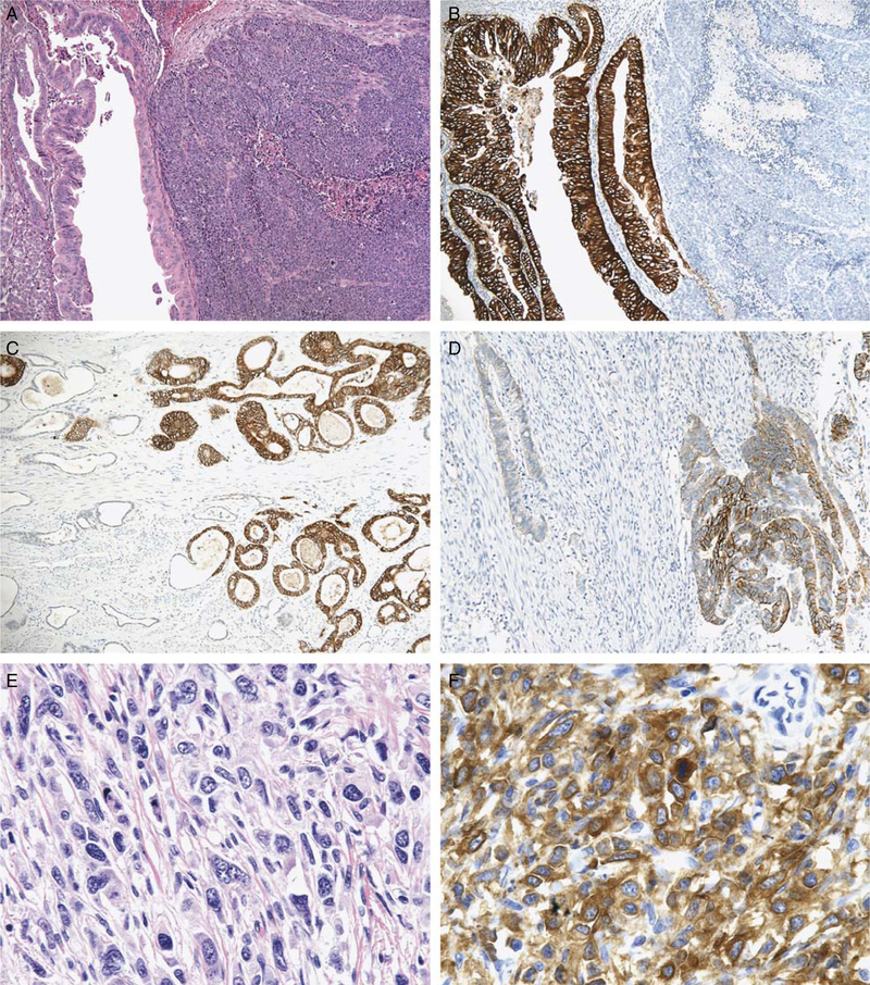
FIGURE 2.
Gastroesophageal intestinal-type adenocarcinoma demonstrating heterogeneous morphology with intermixed glandular (left half of image) and solid (right half of image) growth patterns [A;hematoxylin and eosin (H&E, × 100)]. The glandular component displayed 3+ HER2 protein overexpression, whereas the solid component lacks HER2 protein expression (B, × 100). Gastric intestinal-type adenocarcinoma demonstrating heterogeneous HER2 protein overexpression with areas exhibiting 3+ expression (right half of image) adjacent to tumor glands lacking HER2 expression (left half of image) (C, × 100). This gastric intestinal-type adenocarcinoma with HER2 gene amplification (not shown) demonstrated strong HER2 protein expression in only 10% to 30% of tumor cells (D, × 200). Gastric diffuse-type adenocarcinoma (E, H&E, × 400) demonstrating cytoplasmic HER2 protein expression in the tissue microarray analysis (F, × 400) and HER2 gene amplification by fluorescence in situ hybridization (not shown).

