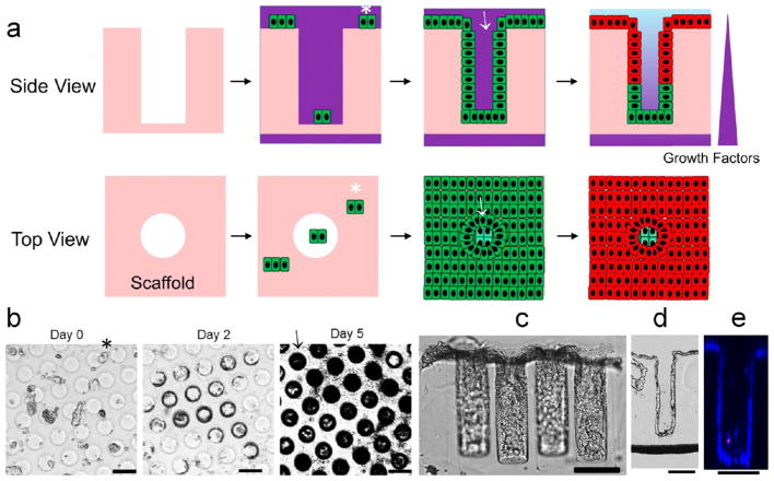
Figure 5.
Strategy to fold and polarize the monolayer of colonic epithelial cells. (a) Schematic showing how the microwell scaffold guides the folding of the cell monolayer into a cryptlike geometry, followed by application of a chemical gradient to polarize the crypt. Top row: side views of a slice through the array. Bottom row: top views looking down onto the array and into the crypts. Fragments of a precultured monolayer (indicated with *) were allowed to settle onto the scaffold, attach, proliferate, and spread to cover the entire scaffold surface, including lining the walls of the microwells (indicated by →). (b) Time-lapse images showing the propagation of cells across the surface of the microwell array. Microwells appeared darker in these bright-field images as their walls were lined with cells. (c) Side view of in vitro crypts. (d) Bright-field image of a 10 μm-thick cryosection through an in vitro crypt demonstrating the open lumen and a monolayer of cells covering the microwell wall. (e) A z slice through an in vitro crypt obtained by confocal fluoresecence imaging. The cells were stained with Hoechst 33342 (blue) and propidium iodide (red). Scale bars = 100 μm.

