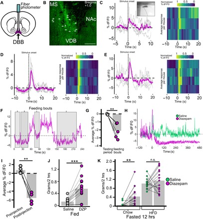
Fig. 4. DBB Vgat neurons are responsive to anxiogenic environmental stimuli, feeding, and diazepam.

(A to E) Response of DBBVgat-GCaMP6m neuronal population to threatening environmental stimuli. (A) Schematic of DBBVgat-GCaMP6m injection and optic fiber placement strategy. (B) Brain slice with DBBVgat-GCaMp6m neurons (VDB); arrows indicate cell bodies. MS, medial septum. (C to E) DBBVgat-GCaMP6m neuronal activity change in response to stimulus (onset at 0 s) with gray traces indicating single interactions, magenta traces indicating averaged response, and heat map indicating normalized average responses per mouse in response to novel object interaction (C; inset shows stimulus onset), looming threat (D), and loud sound (E). (F and G) Response of DBBVgat-GCaMP6m neurons to feeding in the fasted condition with representative trace during feeding bouts (F; shaded gray) and averaged activity during feeding bout compared to average activity over relevant testing period (G; n = 5, P = 0.002). (H to K) Effect of diazepam (DZP) on DBBVgat-GCaMp6 neuronal activity and feeding behavior. (H) Effect of intraperitoneal diazepam on DBBVgat-GCaMp6 neuronal activity in comparison with intraperitoneal saline; (I; n = 4, P = 0.002) Averaged activity 1.5 min before injection compared to average activity 3 to 4.5 min after injection. (J) Effect of intraperitoneal injection of saline versus diazepam on feeding (n = 15) when fed (P < 0.001). (K) Effect of diazepam on fasting-induced food intake of chow and HFD when presented with free access to both simultaneously for 2 hours (chow, P = 0.001; HFD, P = 0.421). *P < 0.05; **P < 0.01; ***P < 0.001.
