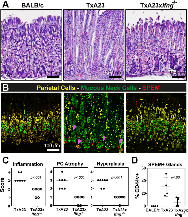
Figure 5. IFN-γ is a critical driver of parietal cell atrophy and SPEM development in vivo.
(A) Representative hematoxylin and eosin stained sections from the gastric corpus of BALB/c, TxA23, and TxA23xIfng−/− mice at 5–7 months of age. (B) Representative fluorescence images showing parietal cells (anti-VEGFB, yellow), mucous neck cells (GS-II, green), and SPEM (anti-CD44v, red). Magenta arrows indicate CD44v+ cells. (C) Scoring of individual stomachs for the degree of inflammatory infiltrate, parietal cell atrophy, and mucosal hyperplasia in TxA23 and TxA23xIfng−/− mice. Each dot represents one mouse, 6–7 mice per group from 2 or 3 separate experiments. (D) Quantification of the fraction of corpus glands containing CD44v+ cells at the base. N=4 mice per group from 2 separate experiments.

