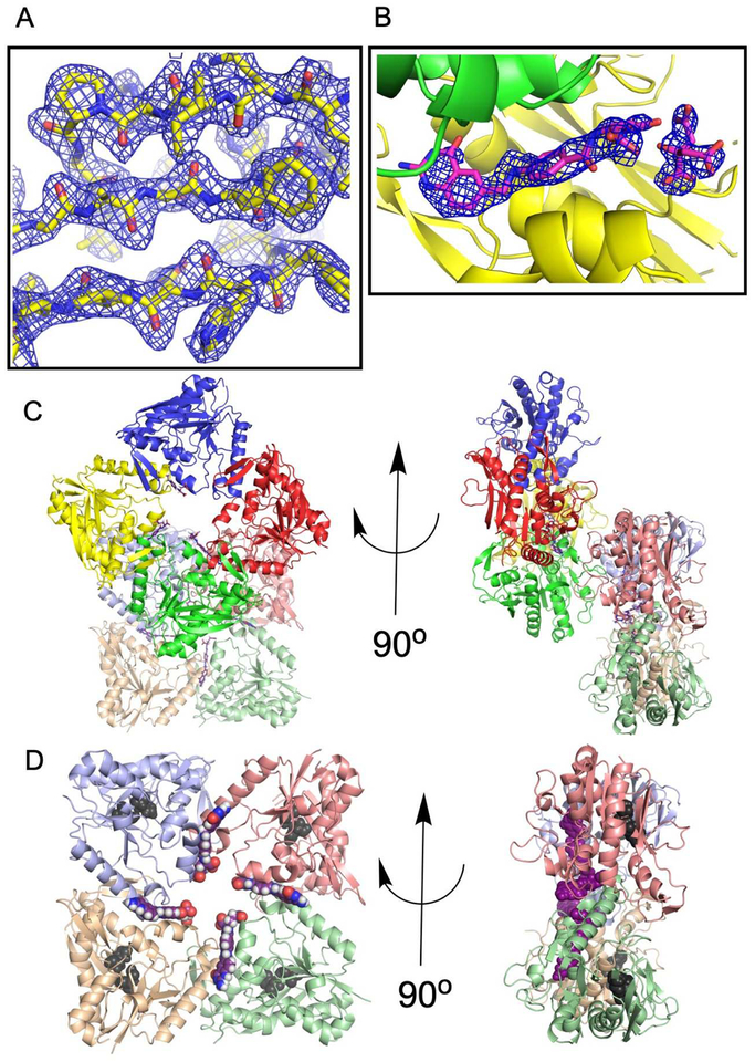
Figure 2.
The asymmetric unit. (A) PvdF crystals appeared single in the drop; however, they showed evident twinning in the diffraction images. Refinement required implementation of the twin law (-h,-k,l). Representative electron density for a 2Fo-Fc simulated annealing map (residues 166–174; 215–243) contoured at 1.5σ is shown. (B) PvdF crystals did not form without the cofactor analogue DDF. Electron density at the interfaces between seven of eight monomers is assigned to DDF and citrate. Electron density is displayed as a 2Fo-Fc simulated annealing omit map contoured at 2σ. (C) PvdF crystallized with eight monomers in the asymmetric unit, as two rings with four-fold symmetry. Each monomer is a distinct color. (D) DDF (magenta) was observed at an interface between PvdF monomers. PvdF monomers E,F,G,H are shown and the location of the active site is highlighted with two of the three residues of the catalytic triad shown in black (the remaining residue is part of a disordered loop). If this were a productive catalytic binding mode, the formate would have to travel ~22 Å. In all other transformylases, the folate binds with the formate directly adjacent to the catalytic triad (within 5 Å).

