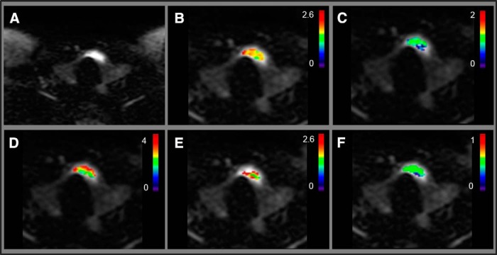
Figure 3.
The representative patient with PTC without tumor-aggressive features (female; 48 years; US maximum tumor diameter, 2.1 cm). Diffusion-weighted image (b = 0 s/mm2) (A). ADC map (×10−3 mm2/s) overlaid on diffusion-weighted image (b = 0 s/mm2) (B). K map overlaid on diffusion-weighted image (b = 0 s/mm2) (C). D* (×10−3 mm2/s) map overlaid on diffusion-weighted image (b = 0 s/mm2) (D). D map (×10−3 mm2/s) overlaid on diffusion-weighted image (b = 0 s/mm2) (E). f map overlaid on diffusion-weighted image (b = 0 s/mm2) (F).

