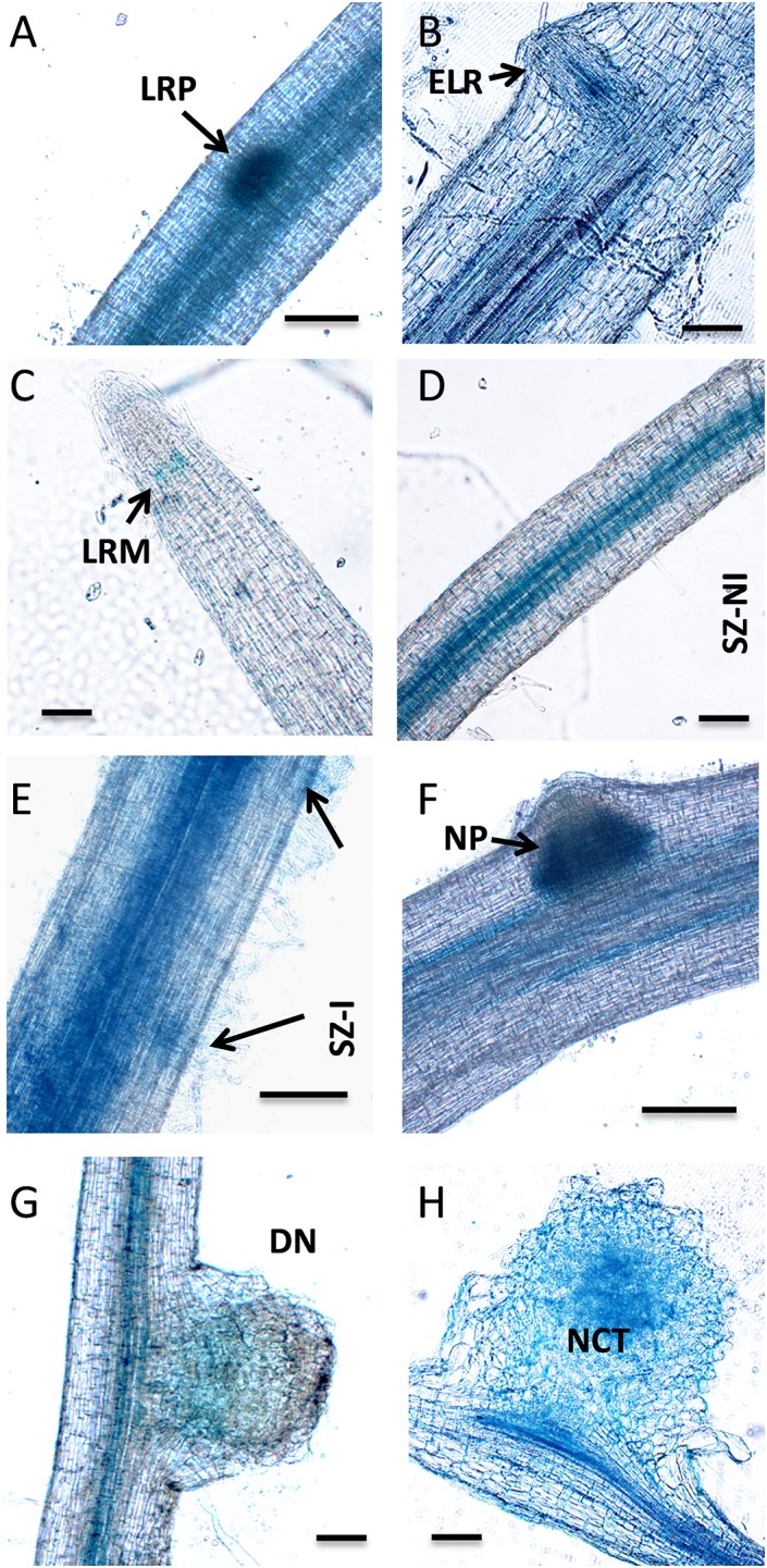
FIGURE 7.

Tissue-specific expression analysis of PvNF-YB7 in roots and nodules of P. vulgaris. Histochemical GUS staining of roots and nodules transformed with the ProPvNF-YB7:GUS construct. (A) Non-inoculated (NI) primary root which containing a lateral root primordia (LRP). (B) Thin section of a primary root containing an emerged lateral root (ELR). (C) Lateral root meristem (LRM) of a NI lateral root. (D) Susceptible zone (SZ) of a NI lateral root. (E) SZ of lateral root inoculated (I) with R. etli strain SC15. Arrows point to infection sites. (F) Nodule primordia (NP) of 6 dpi, (G) developing nodule (DN) of 14 dpi, and (H) mature nodule of 21 dpi formed by strain SC15. NCT: nodule central tissue. Pictures are representative of images observed in more than three independent experiments. Bars: 200 μm.
