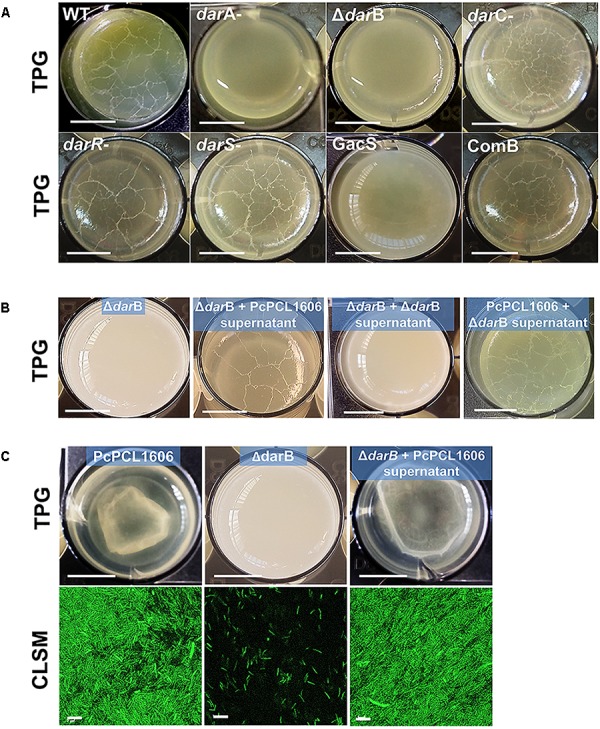
FIGURE 3.

Interphase air–liquid pellicle formation. (A) Pellicle formation of P. chlororaphis PCL1606 and its derivative mutants. Strains were inoculated (final O.D.600nm of 0.08) and grown on TPG media in 24-well plates without agitation at 25°C for 6 days. (B) A similar experiment but using cell-free supernatants of PcPCL1606 mixed with TPG as growth media in order to demonstrate the pellicle recovery in the liquid medium by the defective mutant ΔdarB. (C) The same experiment as B, using GFP-tagged strains to visualize the pellicle using confocal scanning laser microscopy. Size of the bars in the microwell pictures: 5 mm; bars of confocal laser scanning microscopy: 5 μm.
