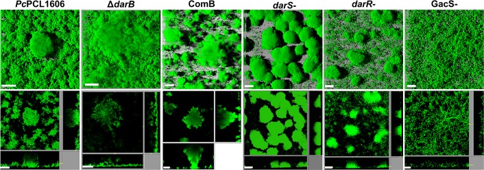
FIGURE 5.
Biofilm architecture of the strains tested. Flow cells were inoculated with a low-density culture of P. chlororaphis PCL1606, the non-HPR producing derivative strain in darB gene (ΔdarB), the complemented ΔdarB derivative strain (ComB), and the transcriptional regulators (ΔdarS, ΔdarR, and GacS) derivative mutants, using AB minimal media supplemented with 1 mM citrate. All the strains were transformed with GFP plasmid for visualization. Biofilm formation was assayed using by confocal laser scanning microscopy. The large frames show the top view, whereas the right and lower frames show vertical sections through the biofilm. Scale bars: 20 μm.

