Fig. 4.
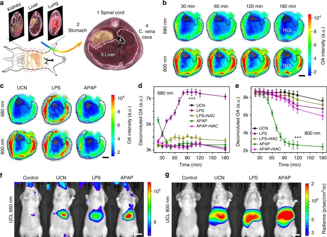
Simultaneous profiling of multiple radicals in inflammation models. a Scheme of multispectral optoacoustic tomography (MSOT) imaging in abdominal regions of live mice (left) and anatomical image of a liver cross-section from the iThea software (right). b Time-resolved MSOT signals at 680 and 800 nm in region of interest (ROI) of liver tomographic images with pseudo-color upon upconversion nanocrystal (UCN) injection (n = 5). c Optoacoustic (OA) images at 680 and 800 nm in ROI of liver upon UCN, lipopolysaccharide (LPS), and acetaminophenol (APAP) administration for 90 min (n = 5). Scale bar: 5 mm. d, e Dynamic profiling of deconvoluted OA signal variations in hepatic inflammation models by UCNs at 680 nm (d) and 800 nm (e) upon LPS, APAP, and their reactive metabolite scavenger (NAC) treatment (n = 5). Statistical significance was assessed by a Student’s t test (heteroscedastic, two-sided). ***p < 0.001. Data were represented as mean ± SD. f, g Representative in vivo upconverted luminescence (UCL) imaging at 660 nm (f) and 800 nm (g) upon saline, UCN, LPS, and APAP treatment for 90 min (Ex: 980 nm, n = 5). Scale bar: 1 cm

