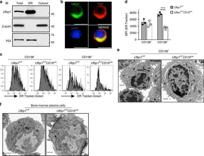
Fig. 4.
Ufbp1 is required for endoplasmic reticulum (ER) expansion in antibody-secreting cells. a Naive B cells from wild-type mice were activated with lipopolysaccharide (LPS) for 3 days. Total, ER, and cytoplasmic fractions were immunoblotted with antibodies against indicated molecules. PDI is used as ER marker, whereas β-actin is a marker for cytoplasm. b Visualization of Ufbp1 and PDI co-localization in LPS-activated B cells by fluorescent microcopy. Scale bars are 5 µm. c Naive B cells from Ufbp1F/F and Ufbp1F/FCD19cre mice were cultured with LPS for 3 days. Shown is the flow cytometric staining of ER-tracker in CD138− and CD138+ cells. d Quantification of ER-tracker staining in CD138− and CD138+ cells in c (n = 3 mice/genotype). MFI mean fluorescent intensity. e Transmission electron microscope images of CD138+ cells sorted from LPS-stimulated B cell cultures from Ufbp1F/F and Ufbp1F/FCD19cre mice. Scale bars are 1 µm. f Transmission electron microscope images of bone marrow plasma cells isolated from mice of indicated genotype. Scale bars are 1 µm. ***P < 0.001. Error bars represent mean ± standard error. Unpaired Student’s two-tailed t-test was used. A representative of at least two experiments is shown

