Fig. 4.
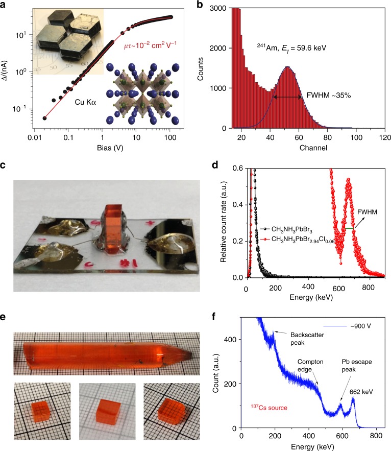
γ-Ray spectrum detection by halide perovskites. a The bias dependence of the photocurrent generated by Cu Kα X-ray in a MAPbI3 single crystal (SC); the red line indicates a fit with the Hecht model. Top inset: Photograph of typical MAPbI3 perovskite SCs grown from a non-aqueous method. Bottom inset: Schematic of the three-dimensional interconnection of PbI6 octahedra in a perovskite lattice (green, Pb; yellow, I; blue, MA). b Energy-resolved spectrum of 241Am recorded with a FAPbI3 SC. c Side view of a CH3NH3PbBr2.94Cl0.06 SC detector, and electrode sides were encapsulated with epoxy. d Enlarged photopeak region of the 137Cs energy spectrum obtained by CH3NH3PbBr2.94Cl0.06 and CH3NH3PbBr3 SC detectors. e As-grown CsPbBr3 SC ingot with a diameter of 11 mm, and the SC wafers with different sizes. f Energy-resolved spectrum of 137Cs γ-ray source with the characteristic energy of 662 keV obtained by a CsPbBr3 detector. a, b (ref. 72), c, d (ref. 46), and e, f (ref. 48) are all adapted with permission from Springer Nature

