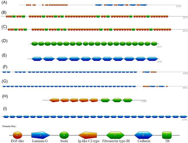
Figure 1.
Domain structures of nine proteins in inherited eye disease. Each panel represents the sequence of domain structures, which varies greatly for each protein, and the domain key can be found at the bottom of the figure. EYS in panel A contains 27 EGF-like (orange circles) and 5 laminin-G domains (blue pentagons). FBN1 and FBN2 in panels B and C, respectively, each contain 47 EGF-like and 9 TB domains (green squares). Panel D shows the 20 sushi domains (green circles) of CFH. Panel E shows the 11 cadherin domains (blue hexagons) of PCDH15. FAT1, in panel F, contains 33 cadherin domains, 5 EGF-like domains and 1 laminin-G domain, while panel G contains the 34 cadherin, 6 EGF-like and 2 laminin-G domains of FAT4. Panel H shows the Ig-like C2-type (orange pentagon) and fibronectin type-III (green hexagon) domains of ROBO3. Panel I show the 27 cadherin domains of CDH23.

