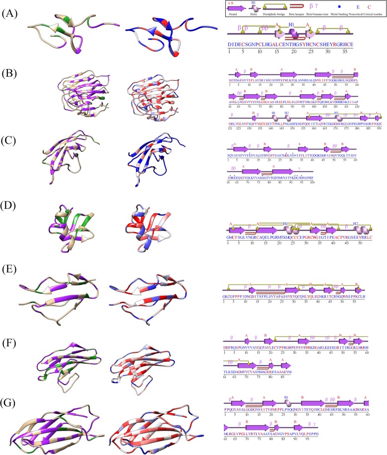
Figure 4.
Domains represented as ribbon structures showing conserved residues (left) foldability of residues (middle), and domain secondary structure components (right). The ribbon structures in the first column highlight nonconserved residues (tan), identically conserved residues (green), and similarly conserved residues (purple). The ribbon structures in the center column are colored by a foldability gradient ranging from blue to white to red; high-foldability residues are in red, and low-foldability residues are in blue. The last column contains secondary structure component plots with motifs and disulfide bonds, as well as the domain sequence with critical residues highlighted in red. The panels show an EGF-like domain of FBN1 (A), a laminin-G domain of EYS (B), a cadherin domain of CDH23 (C), a TB domain of FBN2 (D), a sushi domain of CFH (E), and an Ig-like C2-type (F) and a fibronectin type-III domain of ROBO3 (G).

