Fig. 2.
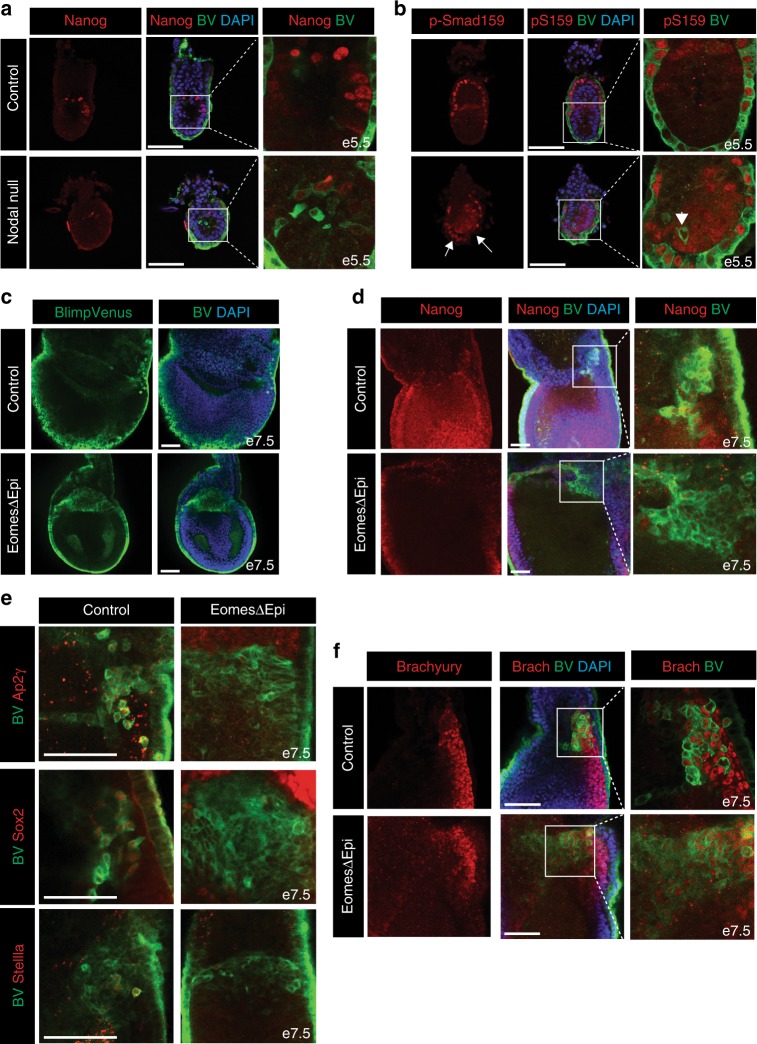
Nodal and its downstream target Eomes regulate early stages of PGC development. a Reduced levels of Nanog expression in e5.5 Nodal null BV+ embryos. b Nodal null BV+ embryos display an expansion of p-Smad159 staining in the distal VE, as indicated by arrows. Arrowhead indicates lowered p-Smad159 staining within BV+ cells. c Expansion of the BV+ cell population in EomesΔEpi embryos at e7.5. d Nuclear Nanog staining in EomesΔEpi and control BV+ embryos at e7.5. e Analysis of Ap2γ, Sox2 and Stella in e7.5 control and EomesΔEpi BV+ cells. f Brachyury staining in control and EomesΔEpi BV+ e7.5 embryos. All IF images are counter stained with DAPI. Scale bars = 100 μm

