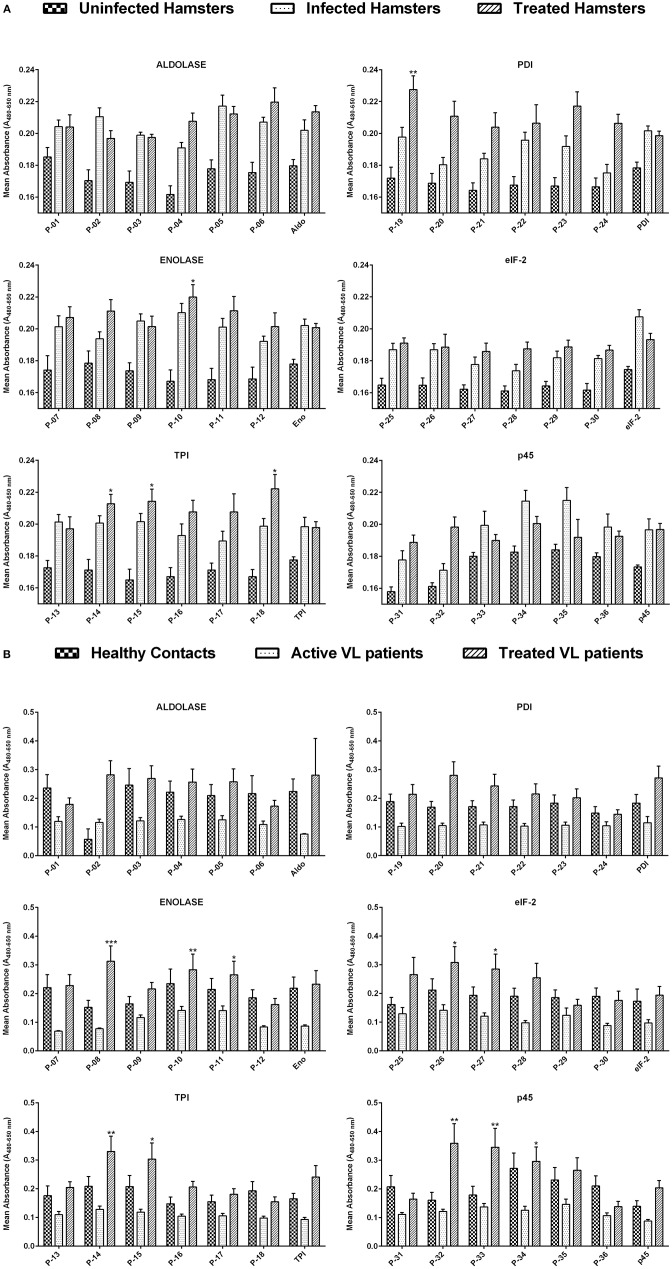
Figure 1.
(A) Lymphoproliferative response in lymphocytes derived from the mesenteric lymph node of uninfected, infected as well as treated Leishmania-infected hamsters against 36 Th1 stimulatory peptides in comparison to their parent recombinant proteins. The concentration of peptides, as well as recombinant proteins, was adjusted to 1 ng/ml of cells. Each bar represents the pooled data of hamsters (Mean ± SE). Statistical significant differences were assessed using a paired t-test amid mean values of treated groups stimulated with synthetic peptides and their respective recombinant proteins. (B) Lymphoproliferative response in PBMCs of healthy contacts, active VL patients as well as treated VL patients against 36 Th1 stimulatory peptides and their parent recombinant proteins. The concentration of peptides, as well as recombinant proteins, was adjusted to 1 ng/ml of cells. Each bar represents the pooled data (Mean ± SE) and the levels of statistical significance were assessed between the mean values of treated groups, stimulated with synthetic peptides and their respective recombinant proteins by paired t-test. *p < 0.05; **p < 0.01; and ***p < 0.001.

