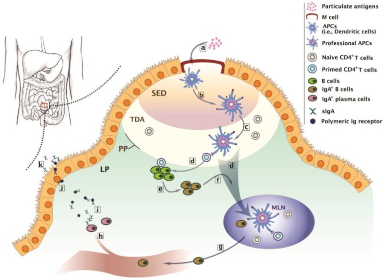
Figure 1.
Biological basis of intestinal immunity at the Peyer’s patches (PP). (a) Transcytosis of particulate antigens to antigen presenting cells (APCs) such as DCs through M cell portal at the inductive sites. (b) Transformation of APCs to professional APCs after antigen-presentation at the subepithelial dome (SED). (c) Priming of naïve CD4+ T cells by professional APCs at the thymus-dependent area (TDA). (d) Activation of B cells by the primed CD4+ T cells, or active migration of professional APCs to the mesenteric lymph node (MLN) for further CD4+ T cell activation and subsequent IgA+ B cell production. (e) Transformation of B cells to IgA+ B cells. (f) Migration of IgA+ B cells to the MLN. (g) The entrance of IgA+ B cells to the systemic circulation through efferent lymph and thoracic ducts. (h) Accumulation of IgA+ B cells at the lamina propria (LP) and maturation of IgA+ B cells to IgA+ plasma cells. (i) The release of dimeric or polymeric IgA from the IgA+ plasma cells. (j) Migration of the complex of dimeric or polymeric IgA with polymeric Ig receptor toward the luminal surface of the intestine. (k) Transcytosis of the complex of and release sIgA at the effector sites.

