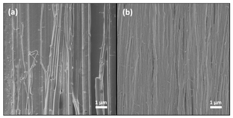
Figure 3.
Scanning electron microscopy (SEM) images of (a) oriented ra-P3HT-TCB and (b) rg-P3HT-TCB films. Both samples show fiber structure in almost only one direction. The ra-P3HT-TCB film has fibers with the diameter from 100 nm to 500 nm. The rg-P3HT-TCB film shows a more uniform distribution of fibers with the diameter of 100–200 nm.

