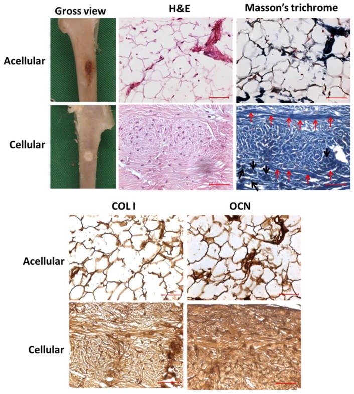
Figure 14.
The gross view, H&E and Masson’s trichrome staining and immunohistochemical (IHC) staining of COL I and OCN of the tibia defects repaired with acellular and cellular hybrid scaffolds 12-week post-operation. The red arrows indicate the osteoid formation while the black arrows denote the existence of osteoblasts in Masson’s trichrome staining for cellular sample. Bar = 50 μm.

