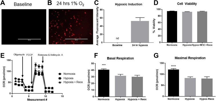
Figure 1.
Cardiomyocyte exposure to 24 hours of 1% oxygen mimics the cellular phenotype of hibernating myocardium (HM). Measurement of cellular hypoxia at (a) baseline and (b) 24 hours following exposure to hypoxic conditions. (c) Quantification of hypoxia by measurement of fluorescent intensity (n=4; p=0.0076; nd=not detectable). (d) Cell viability was unchanged following exposure to 24 hours of hypoxia (n=4). (e) Measurement of oxygen consumption rate (OCR) indicated impaired respiratory capacity following hypoxia that was not restored following reoxygenation. Basal (f) and maximal (g) respiration was significantly reduced following hypoxia and was not restored following reoxygenation (p=0.0005 and p<0.0001, respectively).

