. 2018 Dec 3;8(2):206–215. doi: 10.1039/c8tx00192h

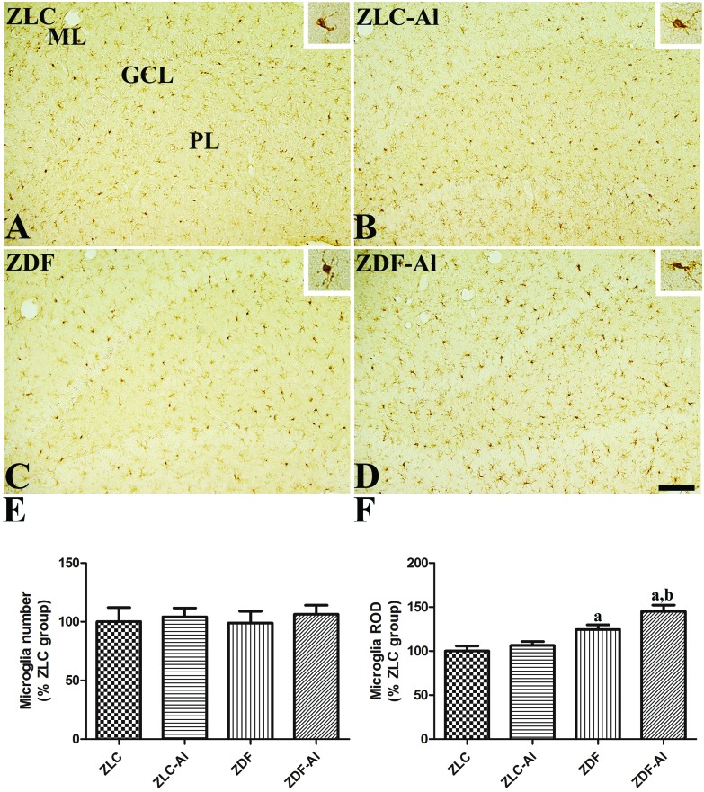
Fig. 4. Ionized calcium-binding adapter molecule 1 (Iba-1) immunoreactivity in the dentate gyrus in ZLC (A), ZLC–Al (B), ZDF (C), and ZDF–Al (D) rats. Iba-1 immunoreactivity is mainly detected in the polymorphic layer (PL) and the molecular layer (ML) of the dentate gyrus in the hippocampus. Iba-1 immunoreactive microglia in ZLC and ZDF rats have less ramified processes than those in ZLC–Al and ZDF–Al rats (inset image). These morphological changes are more prominent in the ZDF–Al group than in the ZLC–Al group. However, there are no significant differences in the number of Iba-1 immunoreactive cells between groups. GCL: granule cell layer. Scale bar = 100 μm. (E) Relative number and the ROD of Iba-1 immunoreactive microglia in the dentate gyrus in the hippocampus are expressed as percentages of the respective values in the ZLC group (n = 5 per group; aP < 0.05 indicates a significant difference between ZLC and ZDF or ZLC–Al and ZDF–Al groups, bP < 0.05 indicates a significant difference between ZLC and ZLC–Al or ZDF and ZDF–Al groups). The error bars indicate the SEM.

Fig. 4