Figure 5.

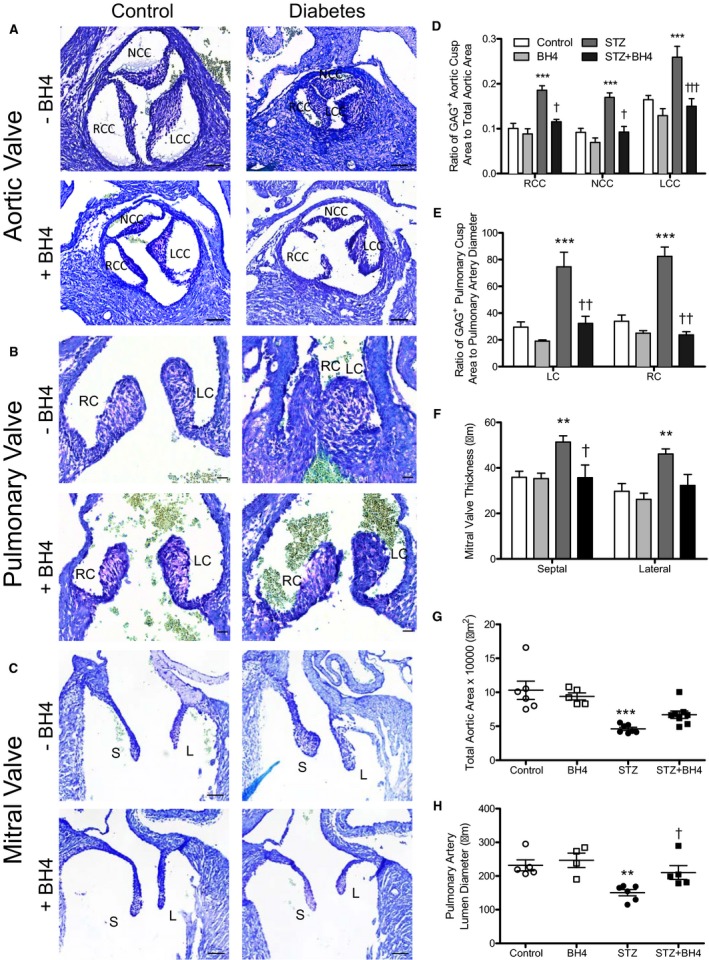
Effects of sapropterin (tetrahydrobiopterin) on aortic, pulmonary, and mitral valve defects induced by pregestational diabetes mellitus at embryonic day (E) 18.5. A through C, Representative images of toluidine blue staining of glycosaminoglycans in aortic, pulmonary, and mitral valves in E18.5 hearts. D, The ratio of glycosaminoglycan‐positive area/total valve leaflet area. E, Pulmonary valve leaflet thickness. F, Mitral valve leaflet thickness. G, Total aortic orifice area. H, Pulmonary artery luminal diameter at the base of the orifice. Bars=50, 20, and 20 μm (A, B, and C, respectively). n=4 to 7 hearts per group from 3 to 4 litters. Data are means±SEM and analyzed using 2‐way ANOVA, followed by Bonferroni post hoc test (B and C). LC indicates left cusp; LCC, left coronary cusp; NCC, noncoronary cusp; RC, right cusp; RCC, right coronary cusp. **P<0.01 vs controls; † P<0.01, †† P<0.01 vs untreated STZ group.
