Figure 1.

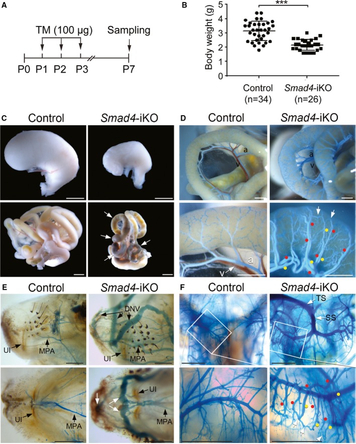
Smad4 deletion results in gastrointestinal hemorrhages and arteriovenous malformations in the intestines, head, and brain. A, Schematic overview of the experimental setup. 100 μg of tamoxifen (TM) was intragastrically injected for 3 consecutive days. B, Decreased body weight of Smad4‐inducible knockout (iKO) pups at postnatal day 7 (P7). All data represent mean±SD. ***P<0.001. C, Morphology of stomachs (upper panels) and intestines (lower panels) of control and Smad4‐iKO pups at P7. Arrows indicate the areas showing hemorrhagic signs. Scale bars: 2 mm. D, Mesenteric and intestinal vessels of controls and Smad4‐iKO visualized by latex dye perfusion via the left ventricle. Red and yellow dots indicate arteries and veins, respectively. White arrows indicate arteriovenous shunting vessels. Scale bars: 1 mm. E, Lateral (upper panels) and bottom‐up (lower panels) views of vasculature in snout areas of control and Smad4‐iKO mice. Apparent arteriovenous shunts were detected in Smad4 mutants. Scale bars: 1 mm. F, Vasculature in the area of the hippocampus of control and Smad4‐iKO visualized by latex dye. Numerous arteriovenous shunts and latex‐filled superior sagittal sinuses were detected in Smad4‐iKOs. Red and yellow dots indicate arteries and veins, respectively. White arrows mark arteriovenous shunts. Scale bars: 1 mm. a indicates artery; DNV, dorsal nasal vein; MPA, major palatine artery; P0, P1, P2, and P3, postnatal day 0, 1, 2, and 3, respectively; SS, straight sinus; TS, transverse sinus; UI, upper incisor; v, vein.
